The retina is a vital component of our visual system. It plays a crucial role in capturing and processing visual information, allowing us to see the world around us. However, like any other part of our body, the retina is susceptible to various disorders that can compromise our vision.
The Anatomy of the Retina
Before delving into the different types of retinal disorders and retinal detachment treatments, it is important to understand the anatomy of the retina. The retina is a layer of tissue located at the back of the eye. It contains specialized cells called photoreceptors, which are responsible for capturing light and converting it into electrical signals that can be interpreted by the brain.
The retina is composed of several key components, each serving a specific function in the visual process.
The Role of the Retina in Vision
One of the primary functions of the retina is to convert light into electrical signals that can be transmitted to the brain via the optic nerve. This allows us to perceive images and interpret the visual world around us. Without a properly functioning retina, our vision would be severely compromised. Visit https://cynergihealth.com/advancements-in-retinal-treatments-new-hope-for-vision-restoration to get about advancements in retinal treatments.

Key Components of the Retina
The retina consists of several layers, each serving a unique purpose in the visual process. The outermost layer is called the pigment epithelium, which absorbs excess light and provides nourishment to the photoreceptor cells.
The layer closest to the pigment epithelium is the photoreceptor layer, which contains two types of photoreceptor cells: cones and rods. Cones are responsible for color vision and high visual acuity, while rods are more sensitive to low-light conditions.
In between the photoreceptor layer and the pigment epithelium lies the retinal pigment epithelium layer, which helps maintain the photoreceptor cells and their function. This layer is crucial for the overall health and integrity of the retina.
Furthermore, within the photoreceptor layer, there are approximately 6 million cones and 120 million rods in the human retina. The cones are densely packed in the central part of the retina, known as the macula, which is responsible for our central vision and detailed visual tasks such as reading and recognizing faces. On the other hand, the rods are more abundant in the peripheral regions of the retina, allowing us to see in dim light and perceive motion.
Lastly, the innermost layer of the retina is made up of various types of nerve cells, including ganglion cells and bipolar cells, which transmit the electrical signals generated by the photoreceptors to the brain. These cells form complex networks that process and refine the visual information before sending it to the brain for interpretation.
Understanding the intricate structure and function of the retina is essential for comprehending the complexities of retinal disorders and their impact on vision. By gaining insight into the different layers and components of the retina, we can appreciate the remarkable precision and coordination required for our visual system to function optimally.
Common Types of Retinal Disorders
Retinal disorders can manifest in various forms, each with its own specific symptoms and treatment options. Here are some of the most common types of retinal disorders:
Retinal Detachment
Retinal detachment occurs when the retina becomes separated from its underlying tissue. This can happen due to trauma, aging, or other underlying medical conditions. Symptoms of retinal detachment include the sudden onset of floaters, flashes of light, and a curtain-like shadow over part of the visual field.
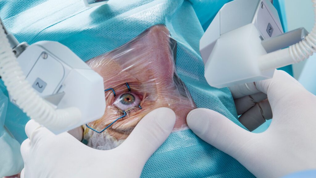
Treatment for retinal detachment often involves surgical intervention to reattach the retina to its original position, preventing further vision loss. During the surgery, the ophthalmologist carefully examines the detached retina and delicately repositions it back into place. They may use tiny sutures or laser therapy to secure the retina in its proper position. After the surgery, patients are closely monitored to ensure proper healing and to address any complications that may arise.
Diabetic Retinopathy
Diabetic retinopathy is a complication of diabetes that affects the blood vessels in the retina. High levels of blood sugar can damage these blood vessels, leading to leakage and the formation of abnormal blood vessels. Symptoms may include blurred vision, dark spots, and floaters.
Treatment options for diabetic retinopathy include laser photocoagulation to seal leaky blood vessels and medication therapies to control blood sugar levels and slow down the progression of the disease. During laser photocoagulation, the ophthalmologist uses a laser to create small burns on the retina, which helps seal off the leaking blood vessels. This procedure is typically performed in an outpatient setting and may require multiple sessions to achieve the desired results.
Macular Degeneration
Macular degeneration is a progressive condition that affects the macula, the central part of the retina responsible for sharp, central vision. There are two forms of macular degeneration: dry and wet. Dry macular degeneration occurs when the macula thins over time, while wet macular degeneration involves the growth of abnormal blood vessels beneath the retina.
Treatment for macular degeneration varies depending on the type and severity of the condition. It may include medication injections, laser therapy, and lifestyle changes such as quitting smoking and consuming a healthy diet rich in antioxidants. Medication injections, such as anti-vascular endothelial growth factor (anti-VEGF) drugs, are administered directly into the eye to inhibit the growth of abnormal blood vessels and reduce inflammation. Laser therapy, on the other hand, uses a focused beam of light to target and destroy abnormal blood vessels, preventing further damage to the macula.
Living with a retinal disorder can be challenging, but with early detection and appropriate treatment, many individuals are able to maintain their vision and lead fulfilling lives. It is crucial to consult with an experienced ophthalmologist who specializes in retinal disorders to receive the most accurate diagnosis and personalized treatment plan.

Symptoms and Diagnosis of Retinal Disorders
Recognizing the signs and symptoms of retinal disorders is crucial for early detection and timely treatment. Here are some common symptoms that may indicate a retinal disorder:
Recognizing the Signs of Retinal Disorders
Blurry or distorted vision, sudden flashes of light, floaters, and a decrease in visual acuity are all common signs of retinal disorders. It is important not to ignore these symptoms and seek professional medical attention as soon as possible.
Retinal disorders can manifest in various ways, affecting the delicate tissue at the back of the eye responsible for capturing light and sending visual signals to the brain. Patients with retinal disorders may also experience a shadow or curtain descending over their field of vision, which could indicate a retinal detachment requiring immediate medical intervention.
Diagnostic Procedures for Retinal Disorders
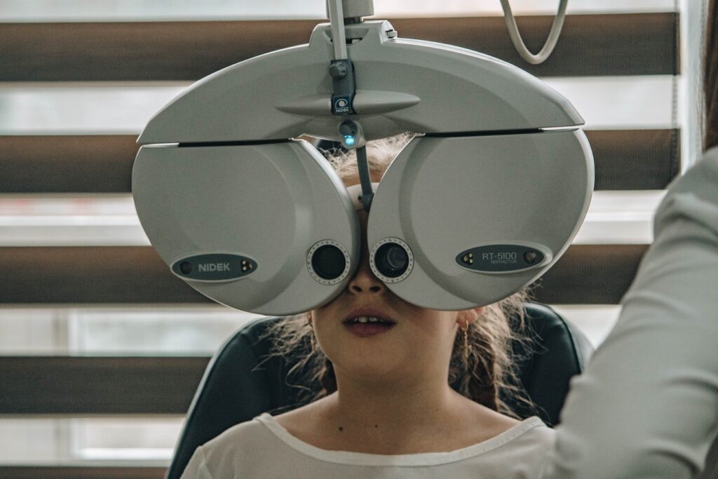
Diagnosing retinal disorders often involves a comprehensive eye examination, which may include visual acuity tests, dilated eye exams, fundus photography, optical coherence tomography (OCT), and fluorescein angiography. These procedures help ophthalmologists assess the health and condition of the retina, guiding further treatment decisions.
Optical coherence tomography (OCT) is a non-invasive imaging test that provides high-resolution cross-sectional images of the retina, allowing for the detection of subtle changes in retinal thickness or the presence of fluid accumulation. Fundus photography, on the other hand, captures detailed color images of the back of the eye, providing valuable information about the structure and integrity of the retina.
Treatment Options for Retinal Disorders
Various treatment options are available for retinal disorders, ranging from surgical interventions to medication and vision rehabilitation. The choice of treatment depends on the type and severity of the retinal disorder.
When it comes to retinal disorders, early detection and prompt treatment are crucial in preventing irreversible vision loss. Regular eye exams are recommended to monitor any changes in retinal health and to initiate appropriate interventions as needed.
Surgical Interventions
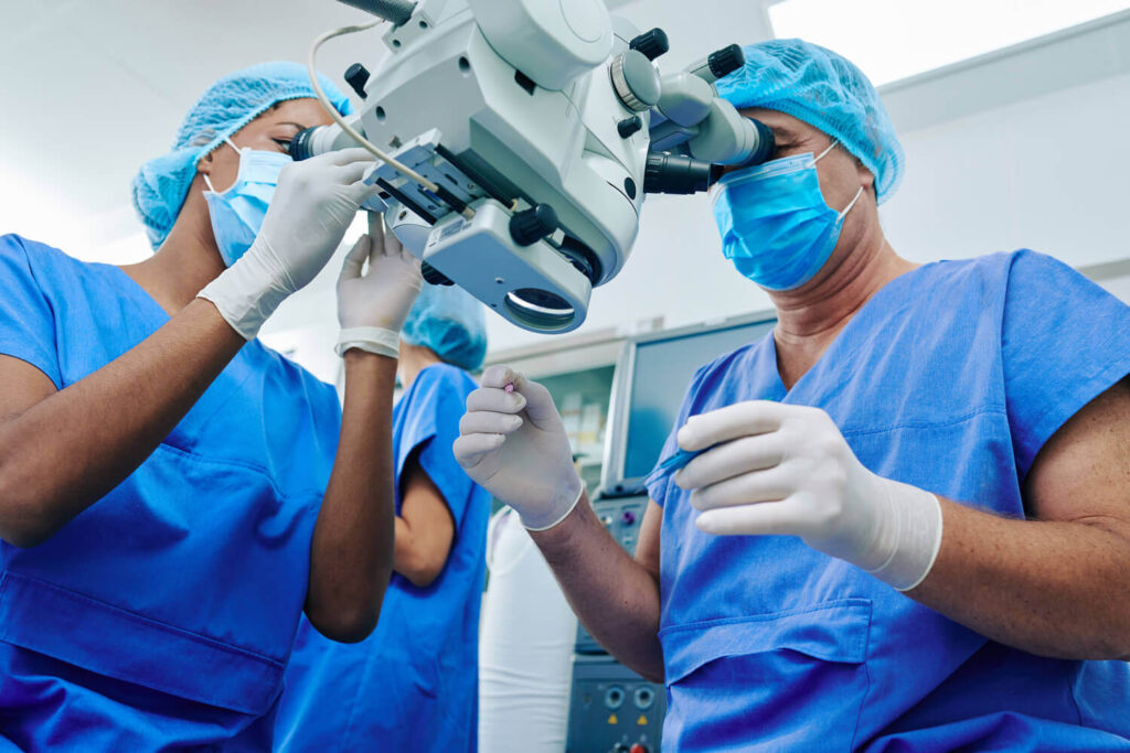
Surgical interventions, such as retinal detachment repair, vitrectomy, and macular hole surgery, can help restore retinal function and improve visual acuity. These procedures aim to reattach or remove damaged retinal tissue, allowing for proper healing and visual recovery.
Advancements in surgical techniques, such as the use of micro-incision vitrectomy systems and intraocular gas injections, have significantly improved surgical outcomes for retinal disorders. These innovative approaches help minimize trauma to the eye and promote faster recovery times for patients.
Medication and Drug Therapies
Medication and drug therapies play a crucial role in treating retinal disorders. Anti-VEGF injections, steroid medications, and immunosuppressive drugs are commonly used to manage conditions such as diabetic retinopathy, macular degeneration, and uveitis. These medications help reduce inflammation, control abnormal blood vessel growth, and preserve retinal function.
Research into novel drug delivery systems, such as sustained-release implants and gene therapies, is ongoing to enhance the efficacy and duration of treatment for retinal disorders. These cutting-edge approaches hold promise for improving patient outcomes and reducing the need for frequent injections or medication adjustments.
Vision Rehabilitation and Support
Vision rehabilitation and support services are essential for individuals with retinal disorders. Visual aids, such as magnifiers and telescopes, can help enhance remaining vision, while low vision rehabilitation programs provide training and support to maximize independence and functionality in daily life.
In addition to traditional vision aids, technological advancements like electronic magnifiers, screen readers, and wearable devices offer new opportunities for individuals with retinal disorders to engage in various activities independently. These assistive technologies continue to evolve, providing tailored solutions to meet the diverse needs of patients with visual impairments.

The Future of Retinal Disorder Treatment
Advances in medical technology and ongoing research hold promise for the future of retinal disorder treatment. Here are some areas of advancement:
Advances in Retinal Surgery
New surgical techniques, including minimally invasive procedures and the integration of robotics, are constantly being developed to enhance surgical outcomes and reduce recovery time. These advancements aim to improve surgical precision and minimize the risk of complications.
Furthermore, the use of advanced imaging technologies like optical coherence tomography (OCT) and adaptive optics has enabled surgeons to visualize the retina with unprecedented detail, allowing for more accurate diagnosis and treatment planning. This level of precision has significantly improved surgical success rates and patient outcomes.
Emerging Drug Therapies
Scientists and researchers are constantly exploring new drug therapies and treatment approaches for retinal disorders. The introduction of gene therapies and neuroprotective agents may hold the key to preventing vision loss and promoting retinal regeneration in the future.
In addition to traditional drug therapies, researchers are investigating the potential of stem cell-based treatments for retinal disorders. Stem cells have the unique ability to differentiate into various retinal cell types, offering a promising avenue for regenerative medicine in the field of ophthalmology.
The Role of Technology in Retinal Disorder Treatment
Technological advancements, such as artificial intelligence-assisted diagnostics, virtual reality rehabilitation programs, and retinal prosthetics, are revolutionizing the field of retinal disorder treatment. These innovative solutions have the potential to significantly improve visual outcomes and enhance the quality of life for individuals with retinal disorders.
Moreover, the development of implantable devices like retinal prosthetics, such as the Argus II retinal prosthesis, has provided new hope for individuals with advanced retinal degenerative diseases like retinitis pigmentosa. These devices work by bypassing damaged retinal cells and directly stimulating the remaining healthy cells, restoring partial vision and improving overall visual function.
Conclusion
Understanding retinal disorders and the available treatment options is crucial for individuals at risk or already affected by these conditions. Early diagnosis, timely intervention, and proper management can significantly improve visual outcomes and preserve overall eye health. With ongoing advancements in medical technology and research, the future looks promising for those affected by retinal disorders.

